Phone
+61 8 8267 2200
Fax
+61 8 8267 5664
Personal Email
In Emergency
0418 829 207
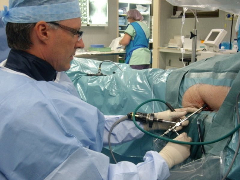
Percutaneous kidney stone surgery (percutaneous nephrolithotomy) is the main method of treatment of larger or complicated upper urinary tract calculi. Urologists concurrently in the UK, USA and Austria developed the original techniques with the first percutaneous kidney stone surgery being performed in Adelaide in 1985. Having been interested in this surgery since assisting at the first case in Adelaide, Denby Steele has been performing percutaneous stone surgery in the supine position since 1999. This previously neglected position is a safer and more efficient approach to the kidney and is gaining increasing popularity around the world. Denby has lectured and run training courses around Australia and overseas on this technique and he has the second largest published series of stones treated this way in the world.
Under general anaesthetic after appropriate positioning with a 3 litre saline bag under the flank of the side with the stone, a cystoscopy is performed where a flexible telescope is passed into the bladder and a hollow catheter is passed up the ureter to the kidney. This is used for injection of radiological contrast to opacify the collecting system to show up on x-ray. Under x-ray control a fine hollow needle is now aimed percutaneously (through the skin) into the kidney as outlined by the contrast material on x-ray. A fine flexible wire is passed through the hollow needle into the kidney, down the ureter and out the urethra. Over this wire a balloon dilator is passed to produce a tract up to about 1cm in diameter over which a hollow plastic sheath is passed to produce a tunnel from outside the body into the kidney through which instruments can be passed to remove the stone or stones. Smaller stones can be extracted with grasping forceps and larger stones will need fragmentation with the holmium laser. A ureteric stent, a narrow flexible hollow polyurethane tube that runs down the ureter with a coil in the bladder at one end and in the kidney at the other end, is usually placed. This serves to allow urine to drain from the kidney down into the bladder around any clots or small stone fragments. A nephrostomy tube, or drainage tube from the kidney exiting the skin through the access site may be left in place but this is uncommon and usually a couple of absorbable sutures are placed to close the puncture. A urethral catheter is left draining the bladder.
If a nephrostomy tube was left, this is usually removed the following morning. If not, the bladdercatheter is usually removed the morning after surgery with a view to discharge that day oncevoiding is established. It is common to experience irritation from the ureteric stent. The small soft coil within thebladder often causes urinary frequency and it is common to experience slight discomfort in thekidney after voiding as urine refluxes up the stent. Haematuria (blood stained urine) is also tobe expected. These symptoms will probably persist while the stent is in place but will be relieved almost immediately the stent several is removedin about a week. The stent is removed by either pulling the fine nylon string left exiting the urethra or returning for a flexible cystoscopy and stent removal. This is performed as a minor procedure through the Day Procedure Unit in hospital without anaesthetic. Though this is frequently the cause of some anxiety, in fact it is not painful either way, only a little uncomfortable for a couple of minutes and nothing to be concerned about.
The above description is for smaller and often solitary stones. Whilst the principles are the same, larger stones and multiple stones can represent quite a challenge. More complex stone surgery can take many hours, sometimes requiring several skin punctures with the chance of needing to return to theatre for a second procedure. Nephrostomy tubes and ureteric stents maybe left in place longer than usual.
Percutaneous surgery is rarely associated with significant complications. Failure to access the kidney percutaneously is possible if the initial needle cannot be guided into a suitable position within the kidney. In this rare occurrence the procedure may need to be rescheduled. Failure to locate all the stones within the kidney is occasionally seen and could require a subsequent procedure. Damage to the kidney or adjacent organs requiring radiological embolization of a bleeding artery, conversion to an open operation or even loss of the kidney is theoretically possible but extremely rare. Prolonged drainage from the nephrostomy site is occasionally seen but rare with a ureteric stent. A degree of intra operative and post-operative bleeding is inevitable but blood transfusionis uncommon except in the more complex cases. Intravenous antibiotics are given intra-operatively and oral antibiotics sometimes started post-operatively so urinary tract infections are rarely seen and of no major consequence.